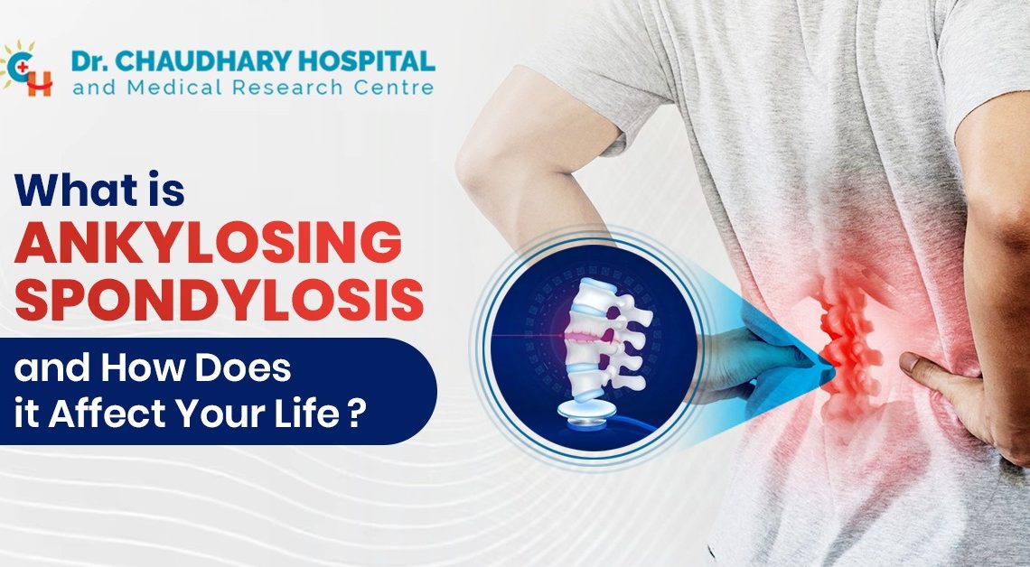
What is Ankylosing Spondylosis and how does it affect your life?

Ankylosing Spondylitis (AS) is not merely a medical term; it’s a life-altering condition that stealthily infiltrates the daily lives of those affected. This chronic inflammatory arthritis primarily targets the spine, leading to pain, stiffness, and, in severe cases, fusion of the vertebrae. In this blog post, we will explore the intricacies of Ankylosing Spondylitis and shed light on how it can significantly impact one’s life.

What is Ankylosing Spondylitis?

Ankylosing Spondylitis is a form of arthritis that predominantly affects the spine, causing inflammation in the spinal joints and ligaments. Over time, this inflammation can lead to the fusion of vertebrae, resulting in a rigid spine. While the exact cause remains unknown, genetic factors are believed to play a crucial role in its development.

Numerous individuals suffering from ankylosing spondylitis experience intermittent back pain and stiffness, ranging from mild episodes to severe, persistent discomfort accompanied by a reduction in spinal flexibility. Furthermore, the impact of the disease extends to other areas of the body, with additional symptoms emerging depending on the specific regions affected. Some individuals may develop associated conditions such as eye disease (uveitis), skin disease (psoriasis), or gastrointestinal issues (inflammatory bowel disease).

While there is no cure for ankylosing spondylitis, various treatment options exist to manage its symptoms effectively. Recommended therapies encompass exercises, along with physical and/or occupational therapy, aiming to enhance mobility and posture. Medications are available to relief pain, control inflammation, improve body position, and slow down the progression of the disease. With proper treatment, the majority of individuals living with ankylosing spondylitis can lead productive lives.

Symptoms and Diagnosis

The symptoms of Ankylosing Spondylitis can vary widely, making it a challenging condition to diagnose. Early signs often include:

  • Pain, stiffness, and inflammation in other joints, such as the ribs, shoulders, knees, or feet.
  • Difficulty taking deep breaths if the joints connecting the ribs are affected.
  • Vision changes and eye pain due to uveitis, which is inflammation of the eye.
  • Fatigue, or feeling very tired.
  • Loss of appetite and weight loss.
  • Skin rashes, in particular psoriasis.
  • Abdominal pain and loose bowel movements.

Diagnosing AS involves a combination of medical history, physical examination, and diagnostic tests such as X-rays and blood tests. However, the journey to a diagnosis can be lengthy, with many individuals facing misdiagnoses and a sense of frustration before receiving a conclusive answer.

The symptoms of ankylosing spondylitis differ among individuals. For some, there are intermittent episodes of mild pain, whereas others experience persistent and intense pain. Whether the symptoms are mild or severe, they may exacerbate during flare-ups and ameliorate during phases of remission.

The Impact on Daily Life

Living with Ankylosing Spondylitis means limitless uncertainty and adapting to a new kind of normal. The unpredictable nature of the condition can make even the simplest tasks a formidable challenge. The stiffness and pain, particularly in the morning, can interfere with daily activities like getting out of bed, tying shoelaces, or even reaching for items on a high shelf.

Beyond the physical challenges, the emotional toll of Ankylosing Spondylitis should not be underestimated. Chronic pain and the potential for disability can lead to anxiety and depression, impacting overall mental well-being. Moreover, the invisible nature of the disease often leads to misunderstandings and skepticism from others, adding a layer of social complexity to the already burdensome experience.

Treatment Approaches

Managing Ankylosing Spondylitis involves a multidimensional approach that combines medication, physical therapy, lifestyle modifications, and, in some cases, surgery. Nonsteroidal anti-inflammatory drugs (NSAIDs) are commonly prescribed to alleviate pain and reduce inflammation. Disease-modifying antirheumatic drugs (DMARDs) and biologics may also be recommended to slow the progression of the disease.

Physical therapy plays a crucial role in maintaining flexibility and preventing deformities. Regular exercise, particularly activities that promote strength and flexibility, such as swimming and yoga, can contribute to improved overall well-being. Additionally, adopting a healthy lifestyle, including proper nutrition and sufficient rest, can positively influence the course of the disease.

The Importance of Awareness

Raising awareness about Ankylosing Spondylitis is important in maintaining understanding and empathy towards the illness. Dispelling misconceptions about the disease and advocating for early diagnosis can contribute to improved outcomes for individuals affected by AS. Education within the medical community and the general public is vital to ensure that those with AS receive timely and appropriate care.

Conclusion

Ankylosing Spondylitis is more than a medical condition; it’s a journey of resilience, adaptation, and self-discovery. Navigating life with AS requires a comprehensive approach that encompasses medical management, lifestyle adjustments, and emotional well-being. By shedding light on the complexities of Ankylosing Spondylitis, we can foster a supportive community that empowers individuals to face the challenges posed by this stealthy foe with courage and grace.

Choosing the Right Surgeon for Your Knee Replacement: What to Look For?

Making the choice to have a knee replacement is a significant step towards living an active life again. It’s critical that you are confident enough in your surgeon. You can increase your chances of a successful procedure and recovery by choosing a surgeon who has performed successful knee replacement surgeries in the past. It’s crucial that you get along well with your surgeon and feel free to ask them any questions or voice any concerns.

Knee replacement is a complex procedure that requires precision and expertise. As a patient, it’s essential to make an informed decision before entrusting your well-being to a particular surgeon. Here are some key factors to consider when choosing the right surgeon for your knee replacement:

Board Certification and Credentials

The first step is to ensure that the surgeon is board-certified and has the necessary credentials and qualifications. Look for certifications from reputable organizations such as the American Board of Orthopaedic Surgery. Additionally, check if the surgeon has received specialized training in knee replacement surgeries and stays updated with the latest advancements in the field.

Experience and Expertise

Assess the surgeon’s experience in performing knee replacement surgeries. Inquire about the number of procedures they have conducted and their success rates. A surgeon with a substantial track record and extensive experience is more likely to handle complex cases and potential complications with expertise and confidence.

Specialization in Knee Surgery

Look for a surgeon who specializes specifically in knee surgeries rather than one who performs a wide range of orthopedic procedures. Specialization indicates a deeper understanding of knee-related issues, which can be critical during the diagnosis, treatment, and post-operative care.

Reputation and Patient Reviews

Research the surgeon’s reputation within the medical community and among past patients. Read reviews and testimonials from other patients who have undergone knee replacement surgeries with the same surgeon. Pay attention to feedback on the surgeon’s communication style, bedside manner, and overall experience during the treatment process.

Hospital Affiliation and Facilities

Check the hospital or surgical center where the surgeon operates. Ensure that the facility is well-equipped with advanced technology and maintains high standards of patient care and safety. A reputable hospital or clinic reflects positively on the surgeon’s commitment to quality care and a conducive surgical environment.

Communication and Trust

Effective communication between you and the surgeon is crucial. During the initial consultation, assess how well the surgeon listens to your concerns and explains the procedure, potential risks, and expected outcomes. A trustworthy surgeon will prioritize your well-being, provide comprehensive information, and encourage open dialogue throughout the treatment journey.

Patient-Centered Approach

Look for a surgeon who adopts a patient-centered approach, considering your unique needs, lifestyle, and expectations. They should be willing to discuss alternative treatment options, if applicable, and involve you in the decision-making process. A surgeon who values patient input is more likely to tailor the treatment plan to your specific requirements and preferences.

Post-operative care and Rehabilitation Support

Inquire about the surgeon’s approach to post-operative care and rehabilitation. A comprehensive follow-up plan and access to physical therapy services are essential for ensuring a successful recovery and maximizing the benefits of knee replacement surgery. A dedicated approach to post-operative care demonstrates the surgeon’s commitment to your long-term well-being.

Surgical Complication Rates and Infection Control Measures

Discuss the surgeon’s approach to minimizing surgical complications and preventing postoperative infections. Inquire about their infection control measures and protocols to ensure a safe surgical environment and reduce the risk of complications during the recovery phase.

Referrals and Second Opinions

Don’t hesitate to seek referrals from your primary care physician or other trusted healthcare professionals. Additionally, consider obtaining a second opinion from another reputable knee replacement surgeon to confirm the diagnosis and treatment plan. Multiple expert opinions can provide you with a more comprehensive understanding of your condition and the available treatment options.

Conclusion

Choosing the right surgeon for your knee replacement is a critical decision that can significantly impact your quality of life. By considering these key factors and conducting thorough research, you can make an informed choice and feel confident in your decision as you prepare for the surgery and the subsequent recovery process. Remember that a successful knee replacement surgery can restore mobility, alleviate pain, and improve your overall well-being, allowing you to resume an active and fulfilling lifestyle.

Skull Base Surgery: What You Need to Know About this Complex Procedure

Skull base surgery, a delicate and intricate procedure, involves accessing the area at the bottom of the skull, which houses critical structures like the brainstem and major blood vessels. This surgical discipline has significantly evolved, incorporating advanced techniques and technologies to address various conditions affecting the skull base. Understanding the nuances and implications of this complex procedure is crucial for patients and their families. This comprehensive guide aims to shed light on the intricacies of skull base surgery, its applications, potential risks, and the advancements that have revolutionized its practice.

In recent years, skull base surgery has emerged as a specialized field, amalgamating the expertise of neurosurgeons, otolaryngologists, and other allied healthcare professionals. By delving into the nuances of this intricate procedure, we can gain a comprehensive understanding of its significance, challenges, and the cutting-edge innovations that have transformed its landscape.

An Overview of Skull Base Surgery

Skull-base surgery primarily targets pathologies that affect the intricate network of nerves, blood vessels, and tissues in the base of the skull. These pathologies can include skull base tumors, vascular malformations, cerebrospinal fluid leaks, and infections. Depending on the nature of the condition, various surgical approaches are adopted, ranging from traditional open surgeries to minimally invasive endoscopic techniques. The selection of the appropriate approach is determined by the location and size of the lesion, as well as the overall health status of the patient.

What is Skull-Base Surgery Used For?

Skull base surgery is used for a spectrum of conditions, including

Skull base tumors

These can encompass benign or malignant growths originating from the bone, soft tissues, or nerves of the skull base. The surgical approach may involve resection, debulking, or targeted therapies, depending on the nature and extent of the tumor.

Cerebrospinal fluid leak

These occur when there is a tear or hole in the membranes that surround the brain and spinal cord, leading to the leakage of cerebrospinal fluid. Surgical intervention is often necessary to repair the defect and prevent potential complications such as meningitis.

Vascular malformations

Abnormalities in the blood vessels at the skull base can pose a significant risk to the patient. Surgical procedures aim to address these malformations and restore normal blood flow, minimizing the risk of neurological deficits or hemorrhage.

Infections

Certain infections, such as osteomyelitis or meningitis, may necessitate surgical intervention to remove infected tissues, drain abscesses, and administer targeted antimicrobial therapy.

Surgical Techniques and Innovations

The field of skull base surgery has witnessed remarkable advancements in surgical techniques and technology, leading to improved patient outcomes and reduced morbidity. Some notable techniques and innovations include:

Endoscopic endonasal surgery

This minimally invasive approach allows surgeons to access the skull base through the nasal cavities, thus avoiding external incisions. It offers enhanced visualization and maneuverability, facilitating the removal of tumors and lesions with minimal disruption to surrounding structures.

Stereotactic radiosurgery

This non-invasive technique delivers precise, high-dose radiation to the target area, effectively treating certain skull base tumors without the need for open surgery. It is particularly beneficial for small tumors or lesions that are challenging to reach using conventional surgical methods.

3D-printing technology

Preoperative planning and the creation of patient-specific models using 3D-printing technology have revolutionized the approach to complex skull base surgeries. Surgeons can simulate the procedure, assess potential challenges, and plan the optimal surgical approach, thus enhancing surgical precision and patient safety.

Intraoperative imaging

The integration of advanced imaging modalities, such as intraoperative MRI and CT, enables real-time visualization during surgery, allowing for immediate assessment of the extent of tumor resection and the identification of any residual lesions, thereby improving the overall surgical accuracy.

Risks and Complications

Despite the remarkable progress in skull base surgery, certain risks and complications remain, emphasizing the need for meticulous preoperative evaluation and comprehensive postoperative care. Potential risks may include:

Damage to critical structures

Given the intricate nature of the skull base, there is a risk of unintentional damage to vital structures such as nerves, blood vessels, and the brain itself, leading to potential neurological deficits or functional impairments.

Cerebrospinal fluid leakage

Inadvertent dural tears during surgery can result in cerebrospinal fluid leaks, which, if not promptly addressed, can lead to complications such as meningitis or intracranial infections.

Postoperative hemorrhage

In some cases, postoperative bleeding may occur, necessitating immediate intervention to control bleeding and prevent hematoma formation, which can exert pressure on the brain and cause neurological deficits.

Surgical site infections

Despite stringent perioperative protocols, the risk of surgical site infections remains, emphasizing the importance of vigilant postoperative care, including the administration of prophylactic antibiotics and meticulous wound management.

Patient Care and Recovery

A crucial aspect of skull base surgery involves comprehensive patient care and a structured rehabilitation program to ensure optimal recovery and a successful return to normal activities. This may include

Postoperative monitoring

Close monitoring of vital signs, neurological function, and wound healing is essential in the immediate postoperative period to identify any signs of complications and ensure timely intervention.

Rehabilitation therapy

Depending on the extent of the surgery and the patient’s preoperative functional status, rehabilitation therapy may be recommended to improve motor and cognitive function, enhance balance and coordination, and promote overall quality of life.

Psychological support

Coping with the emotional and psychological challenges associated with skull base surgery can be overwhelming for patients and their families. Providing access to counseling services and support groups can facilitate emotional well-being and foster a positive outlook during the recovery process.

Conclusion

Skull-base surgery, with its intricate nature and high stakes, represents a remarkable intersection of advanced surgical techniques, technology, and multidisciplinary collaboration. As this field continues to evolve, the emphasis on patient-centric care, precision surgery, and holistic rehabilitation remains paramount. Through continued research, technological innovation, and the collaborative efforts of healthcare professionals, the future of skull base surgery holds the promise of improved outcomes and enhanced quality of life for patients facing complex skull base pathologies.

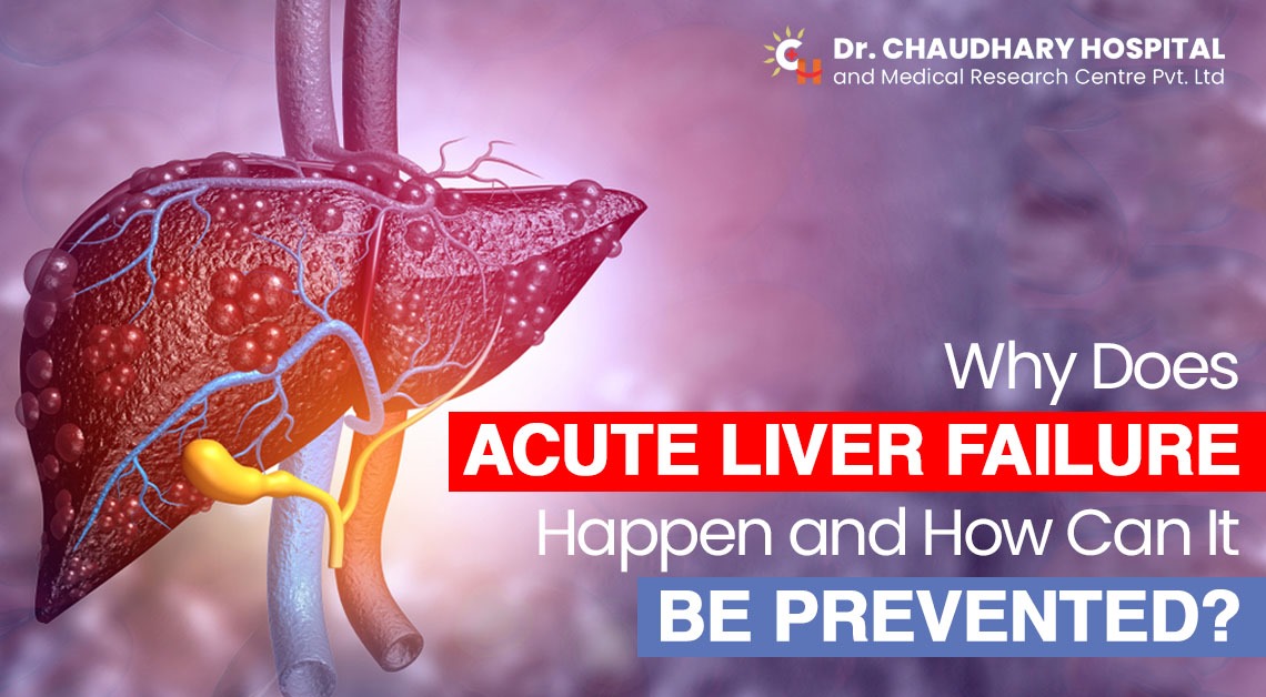
Why Does Acute Liver Failure Happen, And How Can It Be Prevented?

The liver is a vital organ responsible for numerous functions in our body, including detoxification, metabolism, and the production of essential proteins. When the liver suddenly loses its ability to function properly, it results in a life-threatening condition known as acute liver failure (ALF). In this blog, we will delve into the causes of acute liver failure and explore ways to prevent it.

Understanding Acute Liver Failure

Acute liver failure is characterized by the rapid deterioration of liver function within a matter of days or weeks. It can occur suddenly, without any pre-existing liver disease, making it a critical medical emergency. There are various reasons behind this catastrophic event, and understanding its causes is crucial for prevention. Autoimmune disease and Wilson’s disease can also be the leading causes of Acute liver failure.

What Are The Common Causes of Acute Liver Failure?

Viral Infections

Viral hepatitis, especially hepatitis A, B, and E, can lead to acute liver failure. These viruses can attack and damage liver cells, causing a sudden decline in liver function.

Drug-Induced Liver Injury

Certain medications can harm the liver when taken in excessive doses or inappropriately. Acetaminophen (paracetamol) overdose is a notable example, as it can quickly lead to acute liver failure.

Excess Alcohol Consumption

Chronic alcohol consumption can gradually damage the liver, but acute liver failure can occur if a heavy drinker suddenly stops or significantly reduces their alcohol intake.

Autoimmune Hepatitis

In some cases, the immune system mistakenly attacks the liver, leading to inflammation and rapid liver failure.

Wilson’s Disease

This rare genetic disorder causes the accumulation of copper in the liver, leading to acute liver failure if left untreated.

Budd-Chiari Syndrome

This condition occurs when the blood flow out of the liver is obstructed, often due to blood clots in the liver’s veins.

Metabolic Disorders

Certain metabolic diseases, such as acute fatty liver of pregnancy, can trigger acute liver failure during pregnancy as a result the child could also suffer from Jaundice right after birth.

What Are The Symptoms of Acute Liver Failure?

Following are the early signs and symptoms of Acute Liver Failure:

  • The yellow color of the skin and eyes
  • Swollen belly
  • Tiredness & fatigue
  • Extra drowsy and sleepy
  • Nausea
  • Pain in the upper right abdomen
  • A little unwell feeling

How Can You Prevent Acute Liver Failure?

While acute liver failure can be a dire condition, there are steps you can take to reduce your risk and protect your liver health:

Practice Safe Hygiene

To prevent viral hepatitis (hepatitis A, B, and E), maintain good hygiene pracitices, such as washing your hands thoroughly, avoiding consuming contaminated food and water, and getting vaccinated if recommended.

Use Medications Wisely

Always follow your healthcare provider’s instructions when taking medications. Avoid excessive or prolonged use of over-the-counter pain relievers, especially acetaminophen.

Limit Alcohol Consumption

If you choose to drink alcohol, do so in moderation. For those with a history of alcohol abuse, seek help to quit or reduce alcohol intake under medical supervision.

Get Vaccinated

Vaccination against hepatitis B can significantly reduce the risk of viral hepatitis and subsequent liver damage.

Manage Chronic Liver Conditions

If you have a chronic liver condition, such as hepatitis C or fatty liver disease, work closely with your healthcare provider to manage and treat it effectively.

Monitor Your Diet

Maintain a balanced diet rich in fruits, vegetables, and whole grains. Limit the consumption of processed foods, saturated fats, and sugars, as they can contribute to liver damage.

Stay Physically Active

Regular exercise can help control your weight and improve liver health. Aim for at least 150 minutes of moderate-intensity exercise per week.

Avoid Toxins

Be cautious of exposure to toxins and chemicals that can harm the liver, such as industrial solvents and certain cleaning products.

Practice Safe Sex

To prevent hepatitis B and C transmission, practice safe sex by using barrier methods like condoms.

Seek Early Treatment

If you suspect any liver-related symptoms or have risk factors for acute liver failure, seek medical attention promptly. Early diagnosis and intervention can be life-saving.

Conclusion

Acute liver failure is a critical medical condition that can have severe consequences if left untreated. Understanding its causes and taking steps to prevent it is essential for maintaining good liver health. By practicing safe hygiene, using medications responsibly, and adopting a healthy lifestyle, you can significantly reduce your risk of acute liver failure and enjoy a healthier, happier life. Remember, your liver plays a vital role in your overall well-being, so take care of it, and it will take care of you.

What is Black Jaundice and how is it different from Yellow Jaundice?

In recent years, emerging infectious diseases have become a global concern. Among these, the Nipah virus has gained significant attention due to its potential for outbreaks with high mortality rates. Nipah virus infection is a zoonotic disease that can be transmitted from animals to humans. In this blog, we will delve into the symptoms of Nipah virus infection in humans, its risk factors, and preventive measures.

What is Jaundice?

Before we delve into the specifics of black jaundice and yellow jaundice, let’s first understand the common underlying causes and symptoms of jaundice. Jaundice occurs when there is an accumulation of bilirubin in the blood. Bilirubin is a yellow pigment that is formed during the breakdown of haemoglobin in old red blood cells. In a healthy body, bilirubin is processed by the liver, which converts it into a water-soluble form that can be excreted through bile and urine. However, if the liver is not functioning properly or there is an excess production of bilirubin, it can lead to the yellowing of the skin and eyes.

What is Black Jaundice?

Acute liver failure, often known as black jaundice, is a serious medical illness marked by a fast decline in liver function. The term “black jaundice” refers to the more severe form of the disease, whereas “jaundice” often refers to the yellowing of the skin and eyes as a result of elevated bilirubin levels. It necessitates quick medical intervention and care.

A type of jaundice known as Heidan illness (black jaundice) is brought on by persistent and chronic jaundice, frequently accompanied by blood stasis, damp-heat, etc. In Western medicine, the clinical signs of Heldan illness (black jaundice) are comparable to those of cirrhosis, which is brought on by a number of chronic liver conditions. The pathophysiology of Heidan disease (black jaundice), which is typically of the yin jaundice type, is primarily related to blood stasis and dampness stagnation, frequently with damp-heat residue.

What are the symptoms of Black Jaundice?

Jaundice itself is a symptom of an underlying medical issue rather than a standalone condition. The primary symptom of jaundice is the yellowing of the skin and the white part of the eyes (sclera), which occurs due to the accumulation of bilirubin in the body. Bilirubin is a yellow pigment that forms when red blood cells break down.

However, there are some specific symptoms associated with black Jaundice such as follows:

  • Yellowing of the skin and eyes (jaundice)
  • Abdominal pain and discomfort
  • Nausea and vomiting
  • Fatigue and weakness
  • Confusion or altered mental state
  • Dark urine and pale stools
  • Swelling in the abdomen and legs

Conditions such as hepatitis, cirrhosis, gallstones, hemolytic anaemia, and more can lead to jaundice. If you or someone you know is experiencing symptoms of jaundice, it’s important to seek medical attention for a proper diagnosis and appropriate treatment.

What are the Causes and Risk Factors Associated with Black Jaundice?

The following are the causes of Black Jaundice:

  • Viral hepatitis (such as hepatitis A, B, or C)
  • Drug-induced liver injury
  • Alcohol abuse
  • Autoimmune hepatitis
  • Metabolic disorders
  • Wilson’s disease (copper accumulation)
  • Hemochromatosis (iron overload)
  • Infections (like Epstein-Barr virus)
  • Herbal and dietary supplements

It can be more likely to occur if certain risk factors are present, such as a history of liver illness, excessive alcohol consumption, and certain genetic disorders.

Diagnosis of Black Jaundice

The diagnosis of Black Jaundice can involve a measure of steps such as:
  • Assessment and review of preceding medical history
  • Physical examination
  • Blood tests
  • Ultrasound & MRI
  • occasionally liver biopsy

Determining the best course of treatment depends on a timely and accurate diagnosis.

Treatment of Black Jaundice

Medical Attention

Black jaundice episodes require immediate medical care. To handle difficulties and keep the patient stable, hospitalization could be required. Treatment options may include corticosteroids, antiviral drugs, or other particular medicines, depending on the underlying reason.

Lifestyle Changes

Black jaundice patients frequently need to make considerable lifestyle adjustments. This can involve avoiding alcohol, maintaining a healthy weight, and rigorously adhering to medical advice.

Dietary changes

A balanced diet is crucial for supporting liver health. Try to include the following food in your diet:
  • High-fiber foods – fruits, vegetables, whole grains
  • Lean protein sources – chicken, fish, legumes
  • Healthy fats – avocado, nuts, olive oil

Try to avoid the following:

  • Fried and processed foods
  • Excessive salt and sugar
  • Alcohol and high-sugar beverages

What is Yellow Jaundice?

Yellow jaundice, also known as icterus or hyperbilirubinemia, is the most common form of jaundice. It is characterized by the classic yellow discoloration of the skin and eyes. Yellow jaundice can be caused by various factors:

Liver Diseases –

Conditions such as hepatitis, cirrhosis, and liver cancer can impair the liver’s ability to process bilirubin, leading to its accumulation in the bloodstream.

Excessive Hemolysis

Increased breakdown of red blood cells (hemolysis) can overwhelm the liver’s capacity to process bilirubin, leading to its elevation in the blood.

Biliary Obstruction

Blockages in the bile ducts, whether due to gallstones or tumors, can prevent the proper flow of bile and bilirubin, resulting in jaundice.

Conclusion

In conclusion, while yellow jaundice is a well-recognized medical condition characterized by the yellowing of the skin and eyes due to elevated bilirubin levels, black jaundice, or acute liver failure, is a severe condition that demands immediate attention. A medical professional should thoroughly evaluate any discoloration of the skin that appears black, as it could be a call for serious medical treatment.

Black jaundice’s symptoms must be identified, medical attention must be sought promptly, and essential lifestyle adjustments must be made for a successful course of therapy and recovery.

Remember, accurate medical information is crucial for making informed decisions about your health. Always consult reliable sources and healthcare professionals for accurate information and guidance.


What are the Symptoms of Nipah Virus Infection in Humans?

In recent years, emerging infectious diseases have become a global concern. Among these, the Nipah virus has gained significant attention due to its potential for outbreaks with high mortality rates. Nipah virus infection is a zoonotic disease that can be transmitted from animals to humans. In this blog, we will delve into the symptoms of Nipah virus infection in humans, its risk factors, and preventive measures.

What is the Nipah Virus?

A zoonotic virus, the Nipah virus (NiV) transmits from animals to humans. It can also spread through pigs and other animals including goats, horses, dogs, or cats. It is mainly spread by fruit bats, often known as flying foxes. The virus multiplies in the following conditions: 

  • The bodily fluids (blood, excrement, pee, or saliva) of an infected animal come into touch with people or other animals.
  • Foods contaminated by an infected animal are consumed by people.
  • People frequently come into contact with a person who is infected with the Nipah virus while providing care for them.

The Nipah virus was first identified in Malaysia in 1999 during an outbreak that primarily affected pigs and subsequently led to human infections. Since then, Nipah virus outbreaks have been reported in various countries, including Bangladesh, India, and several others in Southeast Asia. Fruit bats, specifically the Pteropus genus, are the natural hosts of this virus.

How Does Nipah Virus Spread?

Humans and animals are the main carriers of the virus. However, it can also pass from one person to another. Caretakers should therefore wear protective gear when administering treatment to someone who has the nipah virus.

The Nipah virus spreads easily. Blood, excrement, urine, and body fluids like saliva can all spread the disease. This means that if you are taking care of someone who has the Nipah virus, you could contract it from their cough or sneeze.

What are the Symptoms of Nipah Virus?

Nipah virus infection can manifest with a range of symptoms, which can make it challenging to diagnose early. The incubation period for the virus is typically between 4 to 14 days, meaning symptoms can appear within this time frame after exposure to the virus. Here are the primary symptoms associated with Nipah virus infection:

  • Fever: The initial symptom is often a high fever, accompanied by flu-like symptoms, including fatigue, weakness, and body aches.
  • Headache: Severe headaches are common in Nipah virus-infected individuals, often making it difficult for them to concentrate or go about their daily activities.
  • Respiratory Distress: As the infection progresses, individuals may experience respiratory distress, including coughing and difficulty breathing.
  • Encephalitis: The Nipah virus is known for its ability to cause encephalitis, which is inflammation of the brain. This can lead to confusion, disorientation, and seizures.
  • Coma: In severe cases, Nipah virus infection can progress rapidly, leading to a coma within a matter of days.
  • Nervous System Involvement: The virus can also affect the nervous system, leading to symptoms such as neck rigidity and muscle tremors.
  • Vomiting and Diarrhea: Gastrointestinal symptoms like vomiting and diarrhea can occur, which can contribute to dehydration and weakness.

What Preventive Measures Can You Take?

Preventing Nipah virus infection primarily involves minimizing exposure to the virus. Here are some essential preventive measures:

Avoid Contact with Infected Animals

Stay away from sick animals, especially pigs and bats, in areas with known Nipah virus outbreaks. Farmers and those working with animals should use protective gear.

Practice Good Hygiene

Wash your hands regularly with soap and water, especially after handling animals or their products. Hand sanitizers can be used when soap and water are not available.

Isolation and Quarantine

Isolate individuals suspected of Nipah virus infection and follow proper infection control practices, especially in healthcare settings.

Educate and Raise Awareness

Communities in at-risk areas should be educated about the virus and its transmission, along with the importance of reporting sick animals and individuals.

Conclusion

Nipah virus infection is a potentially deadly zoonotic disease with a range of symptoms that can make early diagnosis challenging. Understanding the symptoms and risk factors associated with Nipah virus infection is essential for both individuals and healthcare professionals. Preventive measures, such as avoiding contact with infected animals and practicing good hygiene, are key to reducing the risk of transmission. Additionally, ongoing research and public health efforts are vital in the fight against this emerging infectious disease, ensuring a safer future for all.

An introduction to Marburg virus: what it is, where it is found, and how it is transmitted

Both humans and non-human primates can be afflicted by the uncommon but severe hemorrhagic fever or disease known as Marburg virus disease (MVD). The disease is a rare and highly infectious virus. It belongs to the same family of viruses as the Ebola virus and is one of the most deadly viruses known to mankind. It was first identified in 1967 when outbreaks of a mysterious illness occurred in Marburg and Frankfurt, Germany. Symptoms include fever, headaches, vomiting, diarrhoea, and severe bleeding. There is currently no cure or vaccine for the Marburg virus and it can be fatal in up to 88% of cases.

The Filoviridae family includes both the Marburg and the Ebola viruses (filovirus). Although the two diseases are brought on by distinct viruses, they are clinically identical. These illnesses are rare and have the potential to spread in high-fatality outbreaks.

Transmission of Marburg Virus

Initially, human MVD infection results from prolonged exposure to mines or caves inhabited by Rousettus bat colonies.

Marburg virus is an infectious agent that is spread primarily through contact with the bodily fluids of an infected person. In general, it spreads through direct human-to-human contact. It can also be spread by contact with contaminated objects, such as bedding or clothing. In rare cases, it can be transmitted through the air when someone is in close contact with an infected person. There is no known cure for the Marburg virus, so prevention is key. It is important to practise good hygiene, such as washing hands regularly, avoiding contact with infected persons, and avoiding contact with objects that may be contaminated with the virus.

While attending patients with suspected or proven MVD, healthcare personnel have regularly gotten infected. Close contact with patients has led to this when infection control measures are not carefully followed. More severe disease, quick deterioration, and probably a greater fatality rate is linked to transmission via contaminated injection equipment or through needle-stick wounds.

Direct contact with the body of the deceased person who has initially suffered from the disease also contributes to the transmission of Marburg. As long as the virus is present in a person’s blood, they are contagious.

Symptoms

Marburg virus is a rare and deadly virus that is similar to the Ebola virus. Primary symptoms of the Marburg virus may include fever, headache, chills, weakness, muscle aches, and general malaise. Other secondary symptoms may include nausea, vomiting, abdominal pain, chest pain, sore throat, aching joints, and a rash. In more severe cases, the Marburg virus can cause bleeding from the eyes, nose, and mouth, as well as from other organs. In the most serious cases, the Marburg virus can cause organ failure, shock, and death.

Between five and seven days, many patients experience severe hemorrhagic symptoms, and fatal cases frequently involve haemorrhage. Bleeding from the nose, gums, and vagina frequently accompanies fresh blood in vomit and faeces. At the sites of venepuncture (where intravenous access is established to administer fluids or draw blood samples), spontaneous bleeding can be particularly problematic. Patients have experienced persistently high fevers during the acute stage of the illness. Confusion, irritation, and aggression might result from the involvement of the central nervous system. In the late stages of the illness, there have been sporadic reports of orchitis (inflammation of one or both testicles).

Death in fatal cases often happens 8 to 9 days after the onset of symptoms and is frequently preceded by substantial blood loss.

Prevention and Control

For outbreaks to be successfully controlled, community involvement is essential. Human transmission can be decreased by increasing public awareness of the risk factors for Marburg infection and the protective actions that individuals can take. Note a few actions that one can take:

minimising the possibility of community-wide human-to-human transmission brought on by direct or close contact with infected patients, particularly through their body fluids. Avoid making direct personal contact with Marburg patients.

Marburg-affected areas should work to ensure that the populace is well-informed about both the disease’s characteristics and the necessary outbreak containment measures.

Measures to stop the spread of the outbreak include prompt, safe, and respectful burial of the deceased, tracking the health of those who may have come into contact with a Marburg patient for 21 days, and separating healthy people from sick people to stop the spread of the disease, caring for confirmed patients, and adhering to good hygiene and a clean environment.

lowering the potential for sexual transmission. WHO advises male Marburg virus disease survivors to practise safer sex and hygiene for 12 months following the onset of symptoms or until their semen has tested Marburg virus-free twice.

Conclusion

Marburg virus is a rare, but potentially deadly virus that can cause severe hemorrhagic fever in humans and non-human primates. There is currently no approved vaccine or treatment for the Marburg virus, though research is ongoing. The best way to prevent infection is to avoid contact with infected animals and to practise good hygiene when traveling to areas where the virus is known to be present.

Udaipur Receives COVID-19 vaccine- Things You should know about the Coronavirus Vaccine

The year 2020 has carved its mark in the history of the world. After waiting for a very long time, India is set to begin its first Vaccination drive, with its first phase commencing from 16th January 2021. There are various vaccines amidst the pandemic in the global market like Pfizer, Moderna that are been used for immunization against Coronavirus. India produced two vaccines Covishield and Covaxin, Covishield has been tested and has proven its efficacy.

Statistically speaking, Jaipur has the highest number of active cases with 58,512 cases, following which is Jodhpur with 44,574 active cases. Udaipur has 11,691 active cases, which after the vaccination drive should drop further.

Udaipur’s Vaccination drive

  • In Mid-January Udaipur received its first phase of COVID-19 vaccination, COVISHIELD ahead of the mass vaccination drive starting on 16th January 2020.
  • The Government of India has prioritized the health workers and the frontline workers for the vaccination, following which the vaccine would be administered to high-risk people and the elderly population.
  • Udaipur will have 9 centers for the planned vaccinations as per the city government.
  • There will be three sections to every center: Waiting area, Vaccination Area, Under observation area. The recipients of the vaccination will be observed for 30 minutes after the vaccination to check for any side-effects from the vaccine.
  • Recipients who want to get the vaccination have to register on the COWIN app as directed by the Government of Rajasthan and the city of Udaipur.
  • For emergencies related to Coronavirus, anyone can call the numbers 104 and 108 for immediate assistance.

What are the vaccines for Coronavirus?

vaccines for covid 19
India has produced two vaccines to fight Coronavirus, COVISHIELD, and COVAXIN. Covishield is developed by the Serum Institute of India, while Covaxin is developed by Bharat Biotech., a Biotechnology Company headquartered in Hyderabad.

COVISHIELD

Covishield is a COVID-19 vaccine developed by Serum Institute of India, headquartered in Pune. The vaccine has been approved by DCGI and is under circulation for mass vaccination drives planned ahead this year by the government of India. Covishield is the only vaccine produced by India that has proven efficacy. The vaccine is given to some states while the other states have received Covaxin. The vaccines are not interchangeable.
Udaipur has received 1,00,500 Covishield vaccines last week for the first phase of the vaccination drive.

Who should not take the vaccine?

  • People who are exhibiting active symptoms of SARS-CoV-2 infection.
  • COVID-19 patients who have been given anti-SARS-Cov-2 monoclonal antibodies or convalescent plasma.
  • Critically unwell and hospitalized patients (with or without intensive care) due to any illness.
  • People with a history of platelet disorder, clotting factor deficiency, or coagulopathy should only be given the vaccine after cautioning them.

Can those with certain health conditions be administered the vaccine?

  • People with a past history of COVID-19 infection can be immunized.
  • People with a history of chronic diseases – cardiac, neurological, pulmonary, metabolic, and malignancies – are also included.
  • Patients on immunosuppression and immunodeficiency or HIV can be administered the COVID-19 vaccine.

What are the side-effects?

For Covishield, there are a few recorded mild adverse effects that may or may not happen following immunization:

  • Injection site tenderness;
  • injection site pain; headache;
  • fatigue; myalgia (muscle pain); discomfort;
  • pyrexia (an abnormal elevation of body temperature); chills; and nausea.
  • In such cases, paracetamol can be given, the advisory adds.
  • Very rare events of demyelinating disorders” have been reported following the vaccination, although not too many recurring cases.
  • In any case of adverse effects, please contact the nearest available hospital and get examined.
COVAXIN

Covaxin is considered India’s first Indigenous COVID-19 Vaccine developed by Bharat Biotech in collaboration with the Indian Council of Medical Research (ICMR) – National Institute of Virology (NIV). The vaccine received DCGI approval for Phase I & II of Human Clinical Trials and the trials started across India from July 2020. The vaccine’s efficacy is not yet proven as opposed to Covishield which is proven to have worked against the virus. Before giving anyone Covaxin, they should be informed about the same. It is called mandatory Informed consent, wherein the patient can take a day to think if he wants to be immunized.

Who should take the vaccine?

  • The vaccine should only be administered by the health professionals and frontline health workers who are a part of the mass drive.
  • The vaccine is strictly for the patients affected by Coronavirus, health professionals, and elderly people as per the Government’s prioritization for the drive.
  • The vaccine should only be taken by patients infected with the virus, in this case after the approval of the patient or their family about the unproven efficacy.
  • Pregnant women should not take the vaccination
  • The vaccine is only for people of 18 years and above.

What are the side effects?

The vaccine has only been approved by the Government in the Clinical Trial Mode, which would mean that the recipients would have to sign a consent form. In any events of severe or any side effects, Bharat Biotech would have to compensate for the same.
According to the Centre’s fact sheet, these are the common adverse events after administering Covaxin.

  • Injection site pain
  • Fever and Headache
  • Body ache and Abdominal pain
  • Nausea and Fatigue
  • Dizziness and Sweating
  • Cold and Cough

If any of these symptoms surface, post-vaccination, please contact the authorities for further examination and prevention.

Covid-19 has swept over millions of lives globally through the year 2020 and now 2021, the development of vaccines should lower the risk of the virus by a considerable amount. Hospitals and health workers are at the frontline of this crisis, be sure to thank them for their service, as we get ahead of this virus.


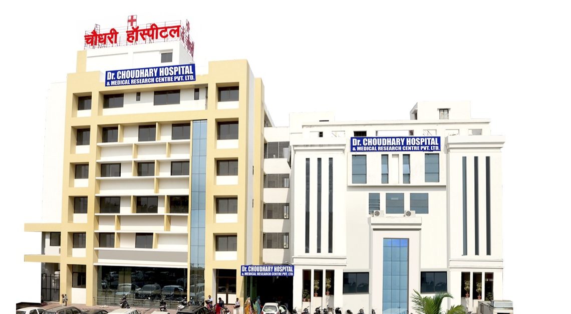
About Chaudhary Hospital

multispeciality hospital in udaipur

Established in 2005, Chaudhary Hospital in Udaipur is the city’s premier Multispecialty hospital situated in the center of the city. Our Hospital is

  • NABH Certified
  • 15 years of experience in Healthcare services
  • 4 Lakh+ tests per year
  • Team of Expert Doctors and Specialists
  • Accurate Results
  • Advanced Diagnostic Labs
  • COVID Care unit

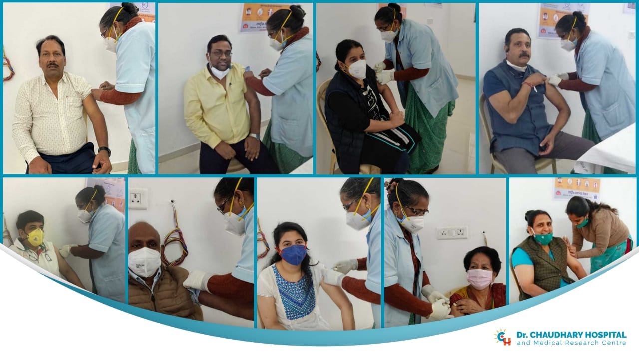
Our team of doctors and the entire staff of Chaudhary Hospital have been vaccinated with the Covishield vaccine. We are proud to have serve Udaipur with the utmost care and safety required to fight the pandemic.

covid vaccination

Our COVID Care unit is equipped with the latest technology and procedures. According to the guidelines by the Ministry of Health and Family Welfare, Government of India, Home Isolation is advisable for entirely asymptomatic or patients with mild symptoms. We at Chaudhary Hospital provide specialized care for COVID-19 infections. To know more, contact us on +91 294 2465566


Understanding HRCT and its Importance in the Diagnostic Process

HRCT means High-Resolution Computed Tomography, it is a cross-sectional imaging technique used for prognosis and diagnosis of various health ailments, most common being lung disease. It is usually performed using a conventional CT Scanner, although the imaging parameters may vary.
The technique of CT Scan was originally very different. There were a lot of complications to be able to get a correct reading, for example, earlier the scans were carried out in wide spaces. The image resolution was an issue. Whereas, the modern CT Scans are very efficient and have a great range of different image resolutions.

When should one get an HRCT Scan?

ct scan

A person should go for an HRCT Scan when:

  • Diagnose muscle disorders, fractured bones, and tumors in bones.
  • To identify sinus problems and other pulmonary disorders
  • To locate exact locations of tumors, blood clots, infections, internal bleeding, or internal injuries.
  • To guide procedures like surgeries, biopsies, radiation therapy.
  • To check for bone diseases
  • To examine structures and regular check-ups of various body organs.

Organs that can be scanned through HRCT Scan are:

  • Chest and Pulmonary CT Scan
  • Thoracic and Lumbosacral Spine CT Scan
  • Cranial CT Scan
  • CT Angiographies
  • CT Enterography

The process of HRCT Scan

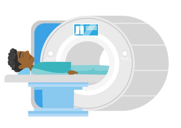
The process of HRCT scans is harmless and pain-free. The patient has to lie face up or down, depending on the procedure on a sliding scan table, that goes into a hollow circular scanner that has cross-sectional high-resolution X-Rays. Sometimes the patient is injected with Dye to outline the organs for a clearer examination of different body parts.

After the Scan

After the HRCT Scan, the patient can resume his activities as per usual. Fasting patients can take their meals regularly. Reports are prepared by the radiologists, the doctors discuss the reports with the patients about the diagnosis. The reports are usually available after 24 hours of the scan.

How is HRCT beneficial in the Diagnosis process of Pulmonary diseases and COVID-19?

covid 19 in the lungs

There are multiple types of Lung diseases, to be able to diagnose the right one, CT Scans are very useful. HRCT examinations provide valuable information that cannot easily be depicted through medical history and other tests. It ensures accuracy in the prognosis or diagnosis of diseases. It determines the progression of the diseases easily and examines all the major and acute symptoms of worsening of diseases.

Many healthcare institutes prefer using the swab method to identify an infection. Although, according to a research study done by the American Journal of Roentgenology, it was concluded that the CT Scan diagnosis of COVID-19 is fairly accurate with an error rate of 4%. Various people have different preferences regarding diagnosis, HRCT has been a primary tool in the early detection of COVID-19.


About Chaudhary Hospital

Established in 2005 by Dr. B.R. Chaudhary, Dr. Chaudhary Hospital is Udaipur’s first NABH Certified multi-specialty hospital. It is one of the top multi-specialty hospitals in the city of Udaipur. Our hospital is renowned and recognized as a premier healthcare provider in Rajasthan. Keeping in mind the ongoing crisis of the COVID Coronavirus outbreak we have a COVID Care ward with quarantine facilities along with world-class Diagnostic Services listed below:

  • 24/7 HRCT Scan
  • Advanced operations theaters
  • Radiology
  • Intense care units
  • Trauma and Emergency
  • Dialysis, Pathology
  • 24/7 Ambulance Service
  • Pharmacy etc.

Contribution by Dr. Chaudhary Hospital to the Healthcare Industry in Udaipur Rajasthan

A hospital is a lifeline for ensuring the health of a nation. Rajasthan is a state with the most amount of tribal and rural population. According to the 1991 census, Udaipur District is one of the poorest districts in the country. The population mostly amounts to tribal populations and rural areas. The healthcare facilities are poor and very few people are literate, among which only 5% of the women population is educated.

After the slow intervention of the healthcare system in the nearby districts along with Udaipur, Dr. BR Chaudhary started this vision of providing healthcare facilities to all the areas nearby to bring about change in the society. He created his first Multispecialty hospital called Chaudhary Hospital in Udaipur. His objective to create a healthcare establishment was to a healthy society. Today has served a large number of people, creating awareness to the most backward societies also.

Contribution of Chaudhary Hospital in transforming the healthcare system of Udaipur

Chaudhary Hospital caters to people from all walks of life and ages, there are various insurance policies in place to help the poor get proper care without money becoming a hurdle. Chaudhary Hospital serves 12 districts of Rajasthan and some parts of Madhya Pradesh. There are-

  • 150+ bedded hospitals
  • Advanced operations theaters
  • Radiology
  • Intense care units
  • Trauma and Emergency
  • Dialysis, Pathology
  • 24/7 Ambulance Service
  • Pharmacy
    Among other diagnostic services. The healthcare facilities range from Pediatric care to internal medicine. Read along as we explore how the hospital became a beacon of hope to various isolated places with a less and poor population with no healthcare facilities in the vicinity or around. It has Performed 30,000+ surgeries, served more than 1,00,000 OPD Patients, Provides multi-specialty services like Medicine, General & laparoscopic Surgery, Paediatric Surgery, Neurology, Neurosurgery, Gastroenterology, Trauma & Orthopaedics, Obstetrics & Gynaecology, Urology, Radiology, Paediatrics, Clinical Oncology, Surgical Oncology, ENT, Pulmonology, Interventional Spine & Pain Management, Dental surgery & Physiotherapy.

Dr. Chaudhary Hospital journey towards excellence

Chaudhary Hospital pioneers in providing world-class care according to the various needs of its patients and surgeons. It is NABH Certified Hospital with COVID Care units and has treated over 1000 COVID Patients. It is best known for being the first multi-specialty hospital in Udaipur. One of the other major USPs of the hospital is its location. The hospital is located in the middle of the city, near the bus station, and at a convenient distance to both of the cities’ railway stations.
The hospital has bridged the gap between people not having any access to healthcare in rural areas to providing healthcare to many underprivileged people and communities. We have hospitals in various rural parts of Rajasthan and Madhya Pradesh.

Esteemed features:

1. Affordable Treatment: Chaudhary Hospital has treatments for all kinds of diseases and ailments. There are various schemes and insurance policies that enable the poor population of the rural areas to be able to cover any expenses that might incur due to expensive medical procedures.

2. Easy access to all facilities: Due to the portable ramps and elevators, mobility is not an issue inside the hospital. Even access to the hospital is very accessible from the bus stops and railway stations. There are various public transportation facilities available to and from the hospital.

3. A larger team of doctors: A Multi-specialty Hospital is the one that has facilities for all ailments and diseases with an expert team of doctors and specialists. The patients are so immensely taken care of with air-conditioned rooms and canteens inside and personalized care. The amenities could include these but not limited to:
General Medicine, General Surgery, Neuro physician, neurosurgeon, gastro physician, gastro surgeon, cardiologist, cardiothoracic surgeon, ENT, pulmonology, ophthalmology, rheumatology, hemato onco, orthopedic, gynecology and obstetric, pediatric, nephrologist, urologist, endocrinologist, plastic surgeon, dentist.

4. Better integration with Insurance Companies: If your insurance company is tied to one specialty hospital or service, they only have coverage for limited treatments and facilities. Those insurance policies cannot have an alliance with all specialty hospitals, therefore if all the facilities and treatments are available under one roof, the insurance companies have better integration with hospitals and have more coverage.

5. Location: Chaudhary Hospital is situated right in the middle of the city in Sector 4, Udaipur. The area is a very busy area and has a footfall of at least 50,000 people daily. The aim of the hospital is to be closer to the Railway Station and the bus station, for the migrants from outside Udaipur, who are seeking treatment, does not have to travel for long to get good healthcare services.
The financial health of a country is very important, keeping in mind the on-going crisis and onset of the recession, there are various things that affect the overall growth of a person and a country, in many ways, they go hand in hand.

Chaudhary Hospital’s Medi claims and Corporate Collaborations

Chaudhary Hospital is affiliated with various recognized Mediclaim companies with collaboration with many good corporate companies, Mediclaim schemes like Bhamashah Swastha Bima Yojna which ensures many underprivileged communities to seek treatment. Companies like Bajaj Allianz, Government of Rajasthan, RIICO, National Insurance, Ajmer Vidyut Nigam Limited, etc. Always striving for providing care to all people successfully, you can avail of privileges and coverage by all our associated firms.

Developed with by nDimensions